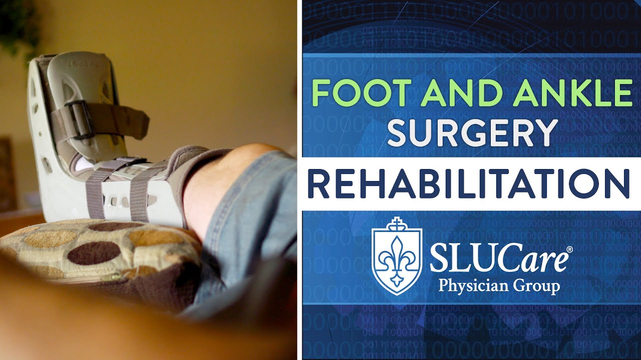
When someone hears the word amputation in a surgical consult, the room goes quiet. The conversation shifts from pain and deformity to independence, balance, and identity. The work of a limb preservation foot surgeon centers on preventing that moment whenever possible. The goal is not just a foot that survives, but a limb that works, bears weight, and fits a life. That means careful diagnosis, thoughtful sequencing of procedures, collaboration across specialties, and relentless attention to biomechanics. It is less about heroics in the operating room and more about strategy over weeks and months.
Over two decades in practice as a foot and ankle surgery doctor, I have learned that preservation is a series of well timed decisions. The patient with a diabetic foot and ankle surgeon NJ midfoot ulcer and Charcot collapse who walks into clinic with a broken custom boot will not be saved by a single operation. The college soccer player with an unstable ankle and cartilage injury will not meet his goals with a brace alone. The grandparent with end stage ankle arthritis who still wants to garden faces a different calculus than the laborer who stands on concrete all day. Each story asks for a plan that protects circulation, fights infection, rebalances forces, and restores alignment.
What limb preservation actually means
Limb preservation is a philosophy that informs a broad set of techniques. At its core, it involves:
- Early control of infection and ischemia, followed by staged reconstruction once biology is stable. Structural correction that redistributes load so wounds close and stay closed. Protection of soft tissue through microinvasive approaches where possible, open approaches when necessary, and plastic surgical coverage when skin is a limiting factor. Realistic functional goals matched to the patient’s life, not a generic notion of normal.
In this work, a foot and ankle preservation surgeon wears many hats. On Monday you may function as a foot and ankle trauma specialist reducing an open calcaneal fracture in a patient with peripheral vascular disease. On Wednesday you may act as a foot and ankle deformity surgeon realigning a Charcot rocker bottom foot. On Friday you could be a foot and ankle wound care surgeon performing a limited debridement on a neuropathic ulcer and applying a biologic dressing. The same week might also include a clinic day as a foot and ankle surgical consultant, reviewing another surgeon’s plan for a complex revision with exposed hardware.
The problems we see most
Diabetic foot ulcers top the list, often complicated by osteomyelitis and poor perfusion. Charcot neuroarthropathy, a destructive neuropathic arthropathy, creates severe midfoot and hindfoot deformity that collapses the arch and shifts pressure to the wrong places. Rheumatoid forefoot deformities, severe bunions with metatarsal malalignment, neglected ankle fractures, posterior tibial tendon dysfunction, and rigid flatfoot all appear frequently. Add to that Achilles tendon ruptures that never healed, repeated ankle sprains with lateral ligament insufficiency, and fractures that united in poor alignment.
A limb preservation foot surgeon also receives referrals after failed operations. These include nonunions after hindfoot fusion, painful or broken implants, or an ankle arthroplasty that loosened. Being a foot and ankle revision specialist means sorting through scar tissue, identifying what went wrong mechanically, and building a safer plan. Sometimes we remove hardware as a foot and ankle hardware removal surgeon to create a healthier landscape before definitive reconstruction.
How judgment drives the sequence
Timing is the overlooked skill. When infection smolders, a foot and ankle emergency surgeon must act quickly. I have performed middle of the night debridements to decompress gas forming infections, remove devitalized tissue, and send reliable cultures. In cases where vascular disease contributes, a vascular colleague handles angioplasty or bypass first. Once the wound bed is granular and perfusion is restored, then a foot and ankle bone realignment surgeon can address the underlying deformity that caused the ulcer.
In athletes with lateral ankle instability and osteochondral lesions, I often start with an arthroscopic procedure as a foot and ankle arthroscopic specialist to address cartilage, combined with ligament stabilization. If peroneal tendons are torn, a foot and ankle tendon surgeon evaluates whether debridement and repair will suffice or if a tendon transfer is needed. Sequencing matters. A clean and well aligned ankle protects cartilage work. In the elderly with valgus ankle arthritis and poor bone density, the decision between joint replacement and fusion requires a candid discussion. A foot and ankle joint replacement surgeon offers motion preservation in the right patient, while a foot and ankle joint fusion specialist may choose a tibiotalocalcaneal fusion for stability when balance and pain outweigh the need for ankle motion.
Diagnostic precision first, then a plan
Good surgery flows from accurate diagnosis. Imaging extends the exam but does not replace hands and eyes. Weight bearing radiographs reveal alignment, collapse under load, and the relationship of joints that non weight bearing films can miss. As a foot and ankle surgical imaging specialist, I lean on CT to map bone loss and subtle malunions, and MRI to assess soft tissue, cartilage, and marrow edema. Ultrasound helps a foot and ankle ultrasound guided surgeon target peritendinous pathology in clinic and direct injections with precision. For nerve entrapments like tarsal tunnel, a foot and ankle nerve decompression surgeon relies on exam findings, Tinel’s sign, and sometimes nerve conduction studies.
Gait analysis refines the plan. As a gait analysis foot surgeon, I study stride length, ankle rocker, pelvic compensation, and pressure mapping. A midfoot ulcer often sits directly under the highest peak pressure on a pedobarograph. Realignment alone may cut that pressure by a third to a half, which can be the difference between a wound that reopens and one that closes for good.
Techniques that protect tissue while fixing structure
The less we disturb healthy tissue, the better it heals. A foot and ankle microinvasive surgeon uses percutaneous screws and small incisions where appropriate, such as for certain osteotomies or calcaneal realignments. An endoscopic approach, in the hands of a foot and ankle endoscopic surgeon, makes sense for plantar fasciotomy or gastrocnemius recession in select patients. A laser assisted foot surgeon may apply laser for adjunctive soft tissue care in resistant warts or nail pathology, though lasers do not replace the fundamentals of wound control and offloading.
Robotics currently play a growing role in alignment planning and, in some centers, in total ankle arthroplasty. A robotic foot and ankle surgeon still relies on tactile judgment, but preoperative CT based guides and intraoperative feedback can improve implant positioning. Navigation reduces outliers. In my practice, those tools have helped keep revision rates low, although the best technology cannot fix poor indications.
When cartilage is salvageable, a foot and ankle cartilage transplant surgeon may use osteochondral plugs or particulated juvenile cartilage. For focal talar dome lesions in younger patients, these options can restore congruency. In degenerative patterns with diffuse loss, a degenerative ankle surgeon counsels patients toward replacement or fusion rather than isolated cartilage work. The skill lies in honest framing of trade offs. A fusion sacrifices motion yet often eliminates pain predictably. A replacement keeps motion and can improve gait but brings risks of loosening. Both can be good choices when matched to the person, the bone, and the demands of daily life.
Bone, tendons, and ligaments as a connected system
Structural correction is rarely one bone cut. A foot and ankle corrective osteotomy surgeon may realign the calcaneus to restore the mechanical axis of the hindfoot, then add a medial column procedure to support the arch. A foot and ankle ligament reconstruction surgeon might stabilize a chronic sprain with a Broström repair, augmented with an internal brace or tendon graft if tissue quality is poor. A foot and ankle tendon transfer surgeon uses the flexor digitorum longus to support a failing posterior tibial tendon, paired with calcaneal osteotomy to shift the heel under the leg.
In certain cavus deformities, peroneus longus to brevis transfer balances the forefoot, while dorsiflexion osteotomy of the first metatarsal lowers a plantarflexed ray and offloads a painful callus. In severe flatfoot with forefoot abduction, lateral column lengthening combined with spring ligament reconstruction restores the medial arch. The foot and ankle biomechanics surgeon’s role is to predict how each cut and each stitch will change load across the plantar surface and the joints.
Wounds, infection, and the long game
The limb preservation foot surgeon must be as comfortable in a wound center as in an operating room. Offloading is non negotiable. Total contact casting remains one of the most effective tools for healing plantar ulcers, often succeeding where expensive dressings fail. After debridement of infected bone, I use antibiotic spacers or beads if the dead space is large, paired with culture directed antibiotics. Once infection is quiet, I plan reconstruction if a deformity persists.
Anecdotally, the fastest healing I have seen in high risk feet came when the team moved in lockstep. The endocrinologist tightened glucose control, the vascular surgeon boosted flow, the wound nurse educated on cast care, and the patient’s family enforced non weight bearing. In those cases, ulcers that lingered for months closed within four to six weeks, and stayed closed after midfoot realignment shifted pressure by measurable amounts.
Regenerative options and when they fit
A foot and ankle regenerative surgery specialist uses tools like platelet rich plasma and stem cell concentrates as adjuncts in specific settings. A PRP foot and ankle surgeon may inject an Achilles tendinopathy refractory to therapy, or apply PRP to a tendon repair to potentially improve healing biology. A stem cell foot surgeon may place bone marrow aspirate concentrate at a nonunion site or within a cartilage defect. These are not magic bullets, and the literature varies. I present them as options, not guarantees, and I reserve them for cases where mechanical alignment is sound and the issue is biology at the margin.
Pediatric and geriatric nuance
Children are not small adults. As a foot and ankle pediatric surgery expert, I watch growth plates and think in years. Flexible flatfoot rarely needs surgery, while rigid deformities from tarsal coalitions may improve dramatically with resection and realignment. In adolescents with osteochondral talar lesions, microfracture or grafting can be successful if we protect the ankle with staged return to sport and careful weight bearing.
The geriatric foot and ankle surgeon faces bone that fractures more easily and skin that tears with tape. Balance and fall risk shape decisions as much as X rays do. After a trimalleolar fracture in an older patient, I may choose hindfoot nails or primary arthrodesis, accepting loss of ankle motion in exchange for earlier protected weight bearing and fewer wound problems. Patients often value predictability over perfect motion.
Acute injuries that threaten the limb
Revascularization and infection control dominate the early hours in open fractures and crush injuries. As a foot and ankle emergency surgeon, I assess skin bridges, zone of injury, and nerve function. Staged protocols help. Initial irrigation and debridement, spanning external fixation to restore length and alignment, and negative pressure dressings buy time for soft tissue to declare itself. Definitive fixation follows once swelling recedes and coverage is secure. In calcaneal fractures with severe blisters, I wait for skin wrinkling to return before incisions, sometimes two weeks. If the soft tissue envelope remains hostile, a foot and ankle joint salvage surgeon may choose percutaneous fixation or a limited sinus tarsi approach to reduce wound complications.
Joint salvage versus replacement
Joint salvage aims to keep a joint functional when possible, using cartilage repair, realignment, or partial fusions to restore more normal mechanics. In the midfoot, limited fusions can eliminate pain at arthritic joints while preserving surrounding motion. In the hindfoot, subtalar or triple arthrodesis restores alignment and stability in neglected deformities. The foot and ankle joint stabilization surgeon’s calculus is mechanical: will the construct bear load without collapsing, and will it prevent recurrence of the deformity that created the ulcer or pain?
Total ankle replacement has matured. A foot and ankle joint surgeon who performs arthroplasty assesses coronal plane deformity, ligament competence, and bone stock. Moderate deformities can be corrected with adjunctive procedures at the time of replacement. In heavier patients or those with neuropathy, I lean toward fusion to avoid catastrophic failure. When a prior replacement fails, a foot and ankle surgical revision expert must decide between revision components and conversion to fusion. I watch the talar body carefully; if it has collapsed, fusion becomes the safer salvage.
Imaging guided accuracy
Targeted accuracy reduces reoperations. As a foot and ankle MRI guided surgeon, I map cysts beneath cartilage, delineate tendon retraction, and spot hidden edema that indicates overload. Needle localization under ultrasound transforms a blind injection into a therapeutic test. Steroid around the peroneal tendons offers a week of relief in tendinopathy. If that window is pain free, surgery focused on the peroneals makes sense. If it is not, I broaden the search. This diagnostic discipline pays off by avoiding surprises in the operating room.
The conversation around risk
No two patients carry the same risk profile. A foot and ankle surgical risk evaluation doctor weighs smoking, HbA1c, vascular studies, neuropathy, BMI, bone density, and the support system at home. If a patient lives alone on the third floor with no elevator and a dog to walk, non weight bearing after a big reconstruction may be unsafe. In that case, I might stage the work or choose a simpler procedure that allows earlier protected weight bearing. Being a foot and ankle outpatient surgery expert and ambulatory surgery specialist means picking cases that can safely go home the same day, with realistic pain control and mobility plans.

Complications still happen. As a foot and ankle surgical complication specialist, I see wound edge necrosis, delayed unions, complex regional pain syndrome, and nerve dysesthesias. Management takes patience. Hyperbaric oxygen sometimes supports healing in compromised tissue. Vitamin D normalization and metabolic bone support help unions. Early recognition of CRPS with desensitization therapy can prevent chronic disability. We talk frankly about probabilities and backup plans.
When appearance matters because function does too
Cosmetic concerns are not frivolous when they impact shoe wear and self perception. A foot and ankle minimally scarring surgeon plans incisions along relaxed skin lines and hides portals. A foot and ankle cosmetic reconstruction surgeon corrects hammer toes and bunions with attention to both alignment and silhouette. Scars that cross joint creases or tether tendons can limit motion. Careful closure matters. When a patient asks for a second opinion on a prior poor cosmetic result that also hurts, the right fix is usually mechanical: straighten the toe, balance the tendons, then close meticulously.
From the first visit to full recovery
Patients often ask what the journey looks like. Although each pathway is personal, the phases are consistent.

- Evaluation and stabilization: history, exam, imaging, vascular and infection workup, pressure mapping, and immediate offloading or bracing as needed. Biological control: debridement if infected, antibiotics when indicated, revascularization if ischemic, wound care until tissue is ready. Mechanical correction: osteotomies, fusions, ligament and tendon procedures, or joint replacement tailored to the problem. Protection and progression: immobilization, staged weight bearing under a foot and ankle weight bearing specialist’s guidance, and targeted therapy to restore gait. Maintenance: custom orthoses, footwear counseling, and periodic surveillance for high risk patients.
Recovery times vary. An ankle fusion typically takes 10 to 14 weeks to unite, with progressive weight bearing starting at 6 to 8 weeks depending on bone quality and fixation. A ligament repair might allow jogging at four months and return to sport between five and eight months. Complex midfoot reconstructions for Charcot deformity may require a frame or cast for months, plus lifetime use of protective footwear.
Red flags that warrant a specialist visit
- A foot ulcer that has not improved after two weeks of proper care or that probes to bone. Deformity that prevents fitting a regular shoe or causes recurrent calluses in the same spot. Recurrent ankle sprains with a feeling of giving way, especially with swelling along the outer ankle. Night or rest pain in the foot or ankle that wakes you and is not explained by a recent injury. A previous foot or ankle surgery that is still painful after the normal healing window or that created new instability.
Tools behind the scenes
Behind every operation sits planning and rehearsal. As a foot and ankle surgical planning specialist, I often create patient specific 3D models for complex malunions. In clinic, as a foot and ankle surgical diagnostics expert, I confirm suspected pathology with targeted exams: squeeze tests for syndesmosis, heel raise for posterior tibial tendon strength, Silfverskiöld test for gastrocnemius tightness. A foot and ankle operative care expert ensures that anesthesia, regional blocks, positioning, and DVT prophylaxis match the case and the patient. After surgery, a foot and ankle surgical recovery expert writes instructions that favor healing, then adjusts them when swelling or activity does not follow the expected curve.
Collaboration that preserves limbs
No limb is preserved in a vacuum. The best outcomes come from team care. The infectious disease physician helps tailor antibiotics, the endocrinologist optimizes metabolic control, the vascular team improves flow, and the plastic surgeon provides coverage when skin is a limiting factor. Physical therapists, pedorthists, and orthotists are essential. As a foot and ankle orthopedic surgical consultant and a foot and ankle podiatric surgical expert, I often bridge disciplines. Titles matter less than shared goals and clear communication.
Why experience changes outcomes
Certain decisions are only obvious in retrospect unless you have seen the pattern before. The painful hardware that looks fine on X ray but sits just proud enough to irritate a tendon sheath. The seemingly minor malalignment that shifts plantar pressure to the ulcer site by a few millimeters, enough to keep it open. The ankle that appears stiff from arthritis but is truly blocked by a tight gastrocnemius. Experience helps a foot and ankle surgical outcomes expert recognize these inflection points. It also teaches humility. I still tell patients when I need more imaging, another opinion, or more time.
Specific scenarios and practical trade offs
A warehouse worker with a painful bunion and a hypermobile first ray wants fast relief. A foot and ankle forefoot specialist might choose a Lapidus fusion to stabilize the medial column. Healing time is longer than a simple distal osteotomy, but recurrence risk drops. If the same patient smokes and cannot take six weeks off work, the equation changes. We discuss a distal procedure with bracing and lifestyle modification, acknowledging a higher chance of recurrence.
A midfoot Charcot patient with a plantar ulcer under the cuboid shows peak pressure of 600 to 700 kPa in that area on gait analysis. After a lateral column lengthening and medial column stabilization by a foot and ankle midfoot specialist, peak pressure falls by half. The ulcer closes in six weeks, and custom shoes maintain the correction.
A runner with posterior tibial tendon pain and a mild planovalgus foot improves with therapy and bracing. When pain persists and imaging shows a partial tear, a foot and ankle tendon surgeon debrides and repairs the tendon, while a foot and ankle alignment surgeon performs a calcaneal osteotomy. She returns to running at six months, with orthoses that support the arch.
The role of second opinions
Complex feet deserve thoughtful review. As a foot and ankle surgical second opinion doctor, I encourage patients to bring prior images, operative reports, and their goals. Sometimes the best move is to continue nonoperative care a bit longer. Other times, a staged approach reduces risk. On more than one occasion, a planned amputation became a salvageable foot once infection was quieted and alignment restored.
Innovation, used wisely
New implants, biologics, and planning tools arrive every year. As a foot and ankle surgical innovations specialist, I trial them cautiously, track outcomes, and discard what does not add value. Small incision bunion techniques promise quicker recovery. In appropriate deformities and with strict attention to three dimensional correction, they can work well. Used indiscriminately, they fail. MRI guided injection techniques improve diagnostic specificity. Robotic guidance in ankle replacement may reduce outliers in component placement. I use innovations in service of fundamentals, not the other way around.
What patients can expect from a preservation mindset
Expect detailed questions about your daily life and your goals. Expect a plan that may include bracing, therapy, offloading, or weight loss before any incision. Expect a conversation about risks that includes your role, from glucose control to smoking cessation to protecting your foot while it heals. Expect options, presented honestly, with enough time to decide. From a foot and ankle clinical surgery specialist you should also expect a team that picks up the phone when problems arise.
The point of saving a limb is not the limb itself. It is the neighborhood walk with your spouse, the job you keep, the grandchild you carry, the sport you love in modified form. A foot and ankle mobility restoration surgeon thinks in those terms. The operative plan and the rehab arc are means to that end.
Preservation succeeds when biology is respected, mechanics are corrected, and the person at the center feels heard. That is the craft, the privilege, and the daily challenge of this work.